
Highlights
- A cystocele occurs when the supportive tissue between the bladder and vaginal wall weakens, often due to childbirth, aging, or chronic pelvic strain.
- Surgical intervention is typically recommended for advanced stages (Stage 3 or 4) or when symptoms like urinary retention and physical discomfort significantly impair quality of life.
- While conservative treatments like pelvic floor exercises and pessaries are first-line options, surgery provides a definitive anatomical repair when non-invasive methods fail.
A cystocele, commonly referred to as a prolapsed bladder, occurs when the supportive fascial tissue between a woman’s bladder and vaginal wall weakens and stretches. While mild pelvic organ prolapse often responds well to non-surgical management, more severe cases can significantly impair daily activities and bladder function. Understanding when does a cystocele need surgery helps women identify critical symptoms and make informed decisions about restoring their pelvic health.
Table of Contents
Understanding Prolapsed Bladder (Cystocele) and Pelvic Support
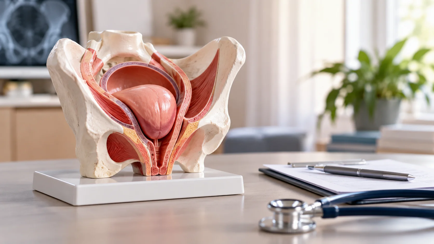
The female pelvic floor consists of a complex network of muscles, ligaments, and connective tissue (fascia) that holds the pelvic organs—including the bladder, uterus, and bowel—in their proper anatomical position. The pubocervical fascia acts as a supportive hammock beneath the bladder. When this fascia tears or stretches, the bladder sags downward, pushing against the anterior (front) wall of the vagina.
Several factors contribute to the weakening of pelvic floor tissue over time:
- Vaginal childbirth: Multiple pregnancies, prolonged pushing, or instrument-assisted deliveries (such as forceps or vacuum extraction) can stretch or tear pelvic muscles and fascia.
- Aging and menopause: Decreased estrogen levels during menopause lead to a loss of collagen and elasticity in pelvic connective tissues.
- Chronic pelvic strain: Persistent coughing from respiratory conditions, repeated heavy lifting, or chronic constipation exerts downward pressure on the pelvic floor.
- Previous pelvic surgery: Hysterectomy or prior prolapse repairs can alter pelvic support dynamics and predispose remaining tissue to laxity.
- Genetic connective tissue weakness: Some individuals naturally possess weaker collagen structures, making them more susceptible to organ prolapse.
In the initial stages, a prolapsed bladder may cause no noticeable symptoms. However, as the protrusion advances, it creates a palpable bulge in the vaginal opening, accompanied by pressure, discomfort, and altered urinary dynamics.
Cystocele Staging: Baden-Walker vs. POP-Q System
To determine the severity of a prolapsed bladder and evaluate whether surgical intervention is appropriate, urogynecologists use standardized staging systems during physical examination.
The Baden-Walker System
Historically, physicians used the Baden-Walker system to grade prolapse based on the extent of descent relative to the hymen:
- Grade 1: The bladder descends halfway to the hymen.
- Grade 2: The bladder descends to the level of the hymen.
- Grade 3: The bladder wall bulges outside the hymen during straining.
- Grade 4: The anterior vaginal wall and bladder fully protrude outside the vaginal opening.
The POP-Q System
Modern urogynecology relies on the Pelvic Organ Prolapse Quantification (POP-Q) system, an objective, reproducible measuring method recommended by international health organizations. POP-Q measures specific anatomical points relative to the hymenal ring in centimeters:
- Stage 0: No prolapse detected; anterior support is fully intact.
- Stage 1: The most distal portion of the prolapse is more than 1 cm above the hymen.
- Stage 2: The prolapse extends between 1 cm above and 1 cm below the hymenal ring.
- Stage 3: The prolapse extends more than 1 cm past the hymen but does not represent complete eversion.
- Stage 4: Complete eversion or protrusion of the anterior vaginal wall outside the body.
Surgical consideration depends heavily on these measurements, but clinical staging alone does not determine the need for an operation. A Stage 2 prolapse in a symptom-free patient may require only observation, whereas a Stage 2 or Stage 3 prolapse causing severe urinary retention requires prompt surgical evaluation.
When Does a Cystocele Need Surgery? Key Clinical Indications
Determining when does a cystocele need surgery involves evaluating anatomical descent alongside the severity of symptoms and the impact on a patient’s overall quality of life. Surgery is rarely urgent, but specific clinical threshold criteria indicate when operative repair is the most effective treatment route.
1. Bothersome Bulge and Physical Discomfort
The most common indication for surgical repair is a persistent, uncomfortable sensation of a bulge or lump at or outside the vaginal opening. Patients frequently describe feeling as though they are “sitting on a ball.” This tissue protrusion can rub against clothing, causing localized irritation, ulceration, and bleeding. When conservative measures fail to relieve this mechanical discomfort, surgery provides physical relief by restoring anatomical alignment.
2. Urinary Retention and Voiding Dysfunction
As the bladder drops below its normal anatomical position, the urethra can become kinked or compressed. This anatomical distortion leads to urinary hesitancy, a weak urinary stream, or the feeling that the bladder never empties completely. Patients often must push against the prolapse (a technique known as “splinting”) to initiate or finish urination. Chronic urinary retention poses serious health risks, including recurrent urinary tract infections (UTIs) and potential kidney damage due to backpressure.
3. Failure or Intolerance of Conservative Treatments
First-line therapy for mild to moderate cystocele includes pelvic floor muscle training (Kegel exercises) and vaginal pessaries. A pessary is a silicone device fitted into the vagina to hold the bladder in place. However, some women cannot tolerate a pessary due to vaginal atrophy, ulceration, discomfort, or difficulty maintaining the device. If pelvic floor therapy fails to control symptoms or a pessary is unfeasible or undesirable, surgical repair becomes the definitive solution.
4. Advanced Stage Prolapse (Stage 3 and Stage 4)
When a cystocele reaches Stage 3 or Stage 4, spontaneous improvement without intervention is extremely unlikely. The extensive fascial rupture prevents conservative therapies from achieving anatomical restoration. In these advanced stages, surgery is indicated to prevent ongoing mucosal injury, resolve severe urinary blockage, and restore pelvic structural integrity.
5. Sexual Dysfunction and Pelvic Pain
Anterior wall prolapse can cause dyspareunia (painful sexual intercourse), excessive vaginal looseness, or fear of tissue damage during intimacy. Additionally, many women experience a constant, heavy dragging sensation in the lower abdomen and back that worsens toward the end of the day or after prolonged standing. When pelvic pain and sexual interference diminish quality of life, surgical correction offers a proven pathway to relief.
Conservative vs. Surgical Management for Cystocele
Choosing between conservative management and operative repair requires comparing non-invasive measures against surgical options.
| Parameter | Conservative Management (PFMT & Pessaries) | Surgical Repair (Anterior Colporrhaphy / Sacrocolpopexy) |
| Primary Goal | Symptom control & progression prevention | Anatomical reconstruction & functional repair |
| POP-Q Suitability | Stage 1 and mild Stage 2 | Moderate Stage 2, Stage 3, and Stage 4 |
| Invasiveness | Non-invasive | Invasive surgical procedure |
| Anatomical Correction | Temporarily supports tissue; does not repair fascia | Reconnects torn fascial layers and elevates bladder |
| Maintenance | Requires continuous exercises or pessary cleaning | One-time procedure with a recovery period |
| Recurrence Risk | Prolapse returns immediately if pessary is removed | Low-to-moderate recurrence risk over long term |
| Ideal Candidate | Mild symptoms, high surgical risk, future pregnancy planned | Severe symptoms, failed pessary, completed childbearing |
Scientific Insights and Research Outcomes
Clinical research published in major urogynecological journals, including practice bulletins from the American College of Obstetricians and Gynecologists (ACOG), establishes that surgical intervention should be symptom-driven rather than based strictly on physical examination findings. Objective clinical trials demonstrate that native tissue anterior colporrhaphy yields subjective cure and symptom relief rates between 68% and 74% over long-term follow-up.Furthermore, advanced minimally invasive techniques—such as laparoscopic or robotic sacrocolpopexy for multi-compartment prolapse—report anatomical success rates exceeding 89%. Research also indicates that surgical correction of severe anterior defects successfully resolves concomitant voiding obstruction and urge urinary symptoms in up to 88% of patients, confirming that restoring anatomical positioning significantly restores lower urinary tract function.
Surgical Procedures for Cystocele Repair
Surgical repair of a prolapsed bladder aims to restore the bladder to its anatomical position and reinforce the supportive pubocervical fascia. The appropriate procedure depends on the patient’s age, sexual activity, previous surgical history, and whether other pelvic organs (such as the uterus or rectum) are also prolapsed.
Anterior Colporrhaphy (Native Tissue Repair)
Anterior colporrhaphy is the standard vaginal procedure for isolated cystocele. The surgeon makes an incision along the front wall of the vagina, folds back the vaginal mucosa, and folds over (plagiarizes) the weakened pubocervical fascia using durable sutures. Excess vaginal epithelium is trimmed, and the incision is closed. This native tissue approach avoids synthetic mesh and offers a smooth recovery with low complication rates.
Paravaginal Defect Repair
When the cystocele is caused by the pubocervical fascia detaching laterally from the pelvic side wall (arcus tendineus fasciae pelvis), a paravaginal defect repair is performed. This approach reattaches the lateral edges of the fascia to the pelvic wall, either through a vaginal incision or via minimally invasive laparoscopy.
Apical Suspension (Sacrocolpopexy or Sacrospinous Ligament Fixation)
A cystocele is frequently accompanied by apical prolapse, where the top of the vagina or uterus descends alongside the anterior wall. Correcting the anterior wall without fixing the apex often leads to surgical failure.
- Laparoscopic / Robotic Sacrocolpopexy: A minimally invasive approach where a surgical mesh strip secures the top of the vagina or cervix to the sacrum (tailbone).
- Sacrospinous Ligament Fixation: A vaginal procedure that anchors the top of the vaginal vault to the strong sacrospinous ligament in the pelvis.
Colpocleisis (Obliterative Surgery)
For elderly or medically frail patients who have severe prolapse and no longer desire vaginal sexual intercourse, colpocleisis offers an effective, low-risk solution. This procedure shortens and closes the vaginal canal, preventing organ protrusion while drastically reducing operative time and anesthesia risks.
Recovery Protocol and Postoperative Care
Following cystocele surgery, proper postoperative care is essential to allow the repaired fascia and sutures to heal without excessive stress.
- Rest and restrict lifting to objects weighing less than 5 kg (10 lbs) for at least 6 to 8 weeks post-surgery.
- Maintain soft bowel movements by consuming high-fiber foods and drinking plenty of fluids to avoid strain during defecation.
- Refrain from vaginal sexual intercourse, tampon use, and douching until cleared by your surgeon at your follow-up exam.
- Perform light daily walking to maintain blood circulation and reduce the risk of deep vein thrombosis.
- Avoid high-impact exercises, squatting, or vigorous physical labor for two to three months following the procedure.
CK Health Turkey: Dedicated Pelvic Floor & Surgical Care
International patients seeking expert medical care for pelvic organ prolapse often look to specialized health destinations that combine surgical excellence with affordability. CK Health Turkey, located in Antalya, provides comprehensive urogynecological and reconstructive pelvic floor care for international patients.
The medical team utilizes advanced diagnostic evaluations and modern, minimally invasive surgical techniques tailored to each patient’s anatomical needs. By offering personalized treatment plans, comfortable recovery settings, and dedicated patient coordination, CK Health Turkey ensures a seamless medical journey for women addressing complex prolapse conditions. Patients interested in discussing surgical options or obtaining a expert medical evaluation can reach out directly or visit the official website to schedule a consultation.
Deciding to undergo pelvic floor reconstruction is a personal medical choice guided by clinical staging, symptom severity, and overall lifestyle impact. Recognizing when does a cystocele need surgery allows women to address urinary dysfunction early, restore pelvic structural integrity, and regain long-term comfort and confidence.