
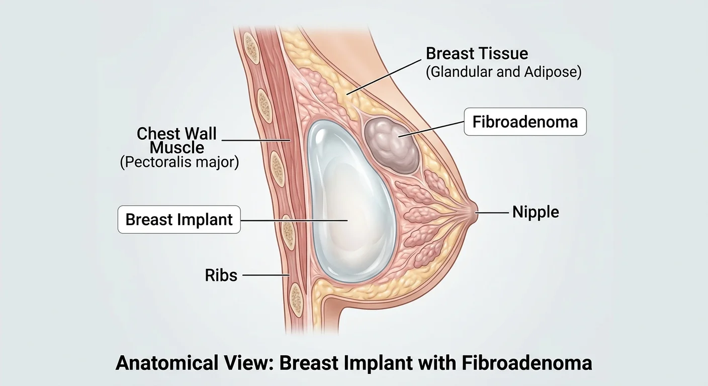
Can you get breast implants with fibroadenoma? In some patients, breast augmentation can still be considered, but the lump must be assessed properly before elective surgery. Whether a fibroadenoma should be monitored, removed first or treated alongside augmentation depends on its diagnosis, size, location and behaviour.
Table of Contents
Can you have breast implants with fibroadenoma?
A confirmed benign fibroadenoma does not automatically make someone unsuitable for breast implants. Fibroadenomas are noncancerous breast growths, and many remain stable or become smaller without treatment. However, a lump that merely seems like a fibroadenoma is not the same as a diagnosis confirmed through an appropriate breast assessment.
Before discussing implant size or incision placement, the clinical team should review your symptoms, examination findings, previous scans and any biopsy reports. If the lump is new, growing or has not been adequately investigated, this needs to be addressed before cosmetic surgery proceeds.
The key question is not simply whether an implant will “fit around” a fibroadenoma. It is whether the breast finding has been diagnosed accurately, whether treatment is indicated independently of augmentation, and how future breast examinations and imaging will be organised.
What is a breast fibroadenoma?
A fibroadenoma is a common, usually well-defined benign breast tumour made of glandular and supporting connective tissue. It often feels like a firm, smooth, moveable lump, although some fibroadenomas are found on ultrasound or mammography without being felt. They are particularly common in younger adults but can occur at other ages.
Fibroadenomas can be single or multiple, and they do not all behave the same way. Some remain unchanged, some become smaller and some grow, including in association with hormonal changes such as pregnancy. Symptoms, size, growth and imaging features all influence whether treatment is recommended.
Breast Cancer Now explains that most fibroadenomas do not need treatment and that simple fibroadenomas are not generally associated with an increased breast cancer risk. A complex fibroadenoma can have additional microscopic features and may be associated with a slightly different risk profile. Your clinician should explain what any pathology result actually means for you.
A fibroadenoma is also different from a fluid-filled cyst and from a phyllodes tumour, another type of fibroepithelial lesion that may need a different surgical approach. This is one reason that imaging and pathology findings must be interpreted together rather than making assumptions based on touch alone.
Why should a fibroadenoma be checked before breast augmentation?
Breast augmentation is elective. It should not delay the investigation of a breast lump that needs assessment. The NHS advises seeking medical assessment for any new breast lump or unusual breast change, even though most lumps are not cancer.
Depending on your age, symptoms and clinical findings, the breast team may recommend an ultrasound, diagnostic mammogram, biopsy or a combination of tests. Ultrasound can help distinguish a solid lesion from a cyst and show its size and position. A core needle biopsy collects tissue for microscopic analysis when it is indicated; not every young patient with a typical imaging appearance needs a biopsy, but uncertain findings must be resolved.
The clinician also checks whether the imaging result, examination and biopsy agree. This is called clinical–radiological–pathological concordance. If a biopsy suggests a fibroadenoma but the scan looks suspicious or the mass behaves unexpectedly, further assessment may still be needed.
Bring any previous ultrasound images, written reports and pathology results to your consultation. A report that identifies a stable, biopsy-proven lesion is much more useful for surgical planning than a vague recollection that a lump was described as “probably harmless”.
Does a fibroadenoma have to be removed before implants?
No universal rule requires every fibroadenoma to be removed before breast augmentation. A small, stable, appropriately evaluated benign lesion may be left in place if the breast specialist considers that safe. Removal can be appropriate when the lump is symptomatic, growing, unusually large, distorting breast contour, uncertain in diagnosis or causing substantial concern after informed discussion.
In a 2025 joint guideline from the American Society of Breast Surgeons and the Society of Breast Imaging, experts advised against routine removal of core biopsy-proven, imaging-concordant fibroadenomas without atypia. They identified symptoms, patient preference, size and substantial growth among the considerations for excision. The same guideline states that these appropriately confirmed uncomplicated lesions do not necessarily require dedicated imaging follow-up beyond age-appropriate screening.
That recommendation applies to correctly assessed fibroadenomas; it is not permission to ignore a new or changing lump. Your surgeon and breast specialist may advise a different plan if the findings are unclear, the lesion is atypical or there is concern for a phyllodes tumour.
Can fibroadenoma removal and breast implants be done together?
In selected patients, a breast surgeon and plastic surgeon may consider fibroadenoma excision and breast augmentation during the same operation. Combining procedures may avoid separate anaesthetic episodes and could allow the surgeons to coordinate incision placement and final breast shape. It is not automatically the safest or best option.
Important considerations include the location and number of lesions, the amount of tissue that would need to be removed, whether the diagnosis is sufficiently certain, infection and bleeding risks, and the possibility that final pathology could require further treatment. If a lesion is suspicious or the diagnosis is unresolved, the priority is a definitive breast assessment, not cosmetic scheduling.
A 2021 small retrospective case series described 10 patients undergoing fibroadenoma excision with breast augmentation and three patients who had fibroadenoma removal after implants were already present. The authors reported that simultaneous surgery was technically possible in their selected patients, but the series was too small to establish general safety or to compare complication rates reliably. It should not be interpreted as proof that combining operations is suitable for everyone.
If the fibroadenoma is removed, the tissue should be sent for appropriate pathological assessment. Surgical teams must also consider how removal could affect breast symmetry, particularly when a sizeable amount of tissue is excised from one side.
Comparing the main options before breast augmentation
| Approach | When it may be considered | Important considerations |
|---|---|---|
| Monitor a confirmed benign fibroadenoma | Stable, appropriately assessed lesion with no reason for removal | Document its baseline findings and follow the breast specialist’s screening or review advice |
| Remove the fibroadenoma before implants | Uncertain diagnosis, significant growth, symptoms, distortion or planned excision | Allows pathology and healing to be reviewed before cosmetic planning; may change breast contour |
| Consider excision and implants together | Selected cases after full breast assessment and multidisciplinary planning | Combines procedures but adds complexity; pathology and potential revision needs must be discussed |
These are discussion pathways rather than treatments a patient can choose without assessment. In particular, a suspicious lesion should not be assumed to be a fibroadenoma or treated as a routine addition to breast augmentation.
Can breast implants make a fibroadenoma grow or turn cancerous?
There is no reliable evidence that placing a breast implant directly causes a benign fibroadenoma to become malignant. Fibroadenoma growth can occur for other reasons and should be evaluated on its own merits. Likewise, an implant does not treat, shrink or remove a fibroadenoma.
The implant and the lump are separate structures: the implant is placed in a surgically created pocket, while the fibroadenoma arises from breast tissue. What matters clinically is the position of the lesion relative to the proposed implant, whether it has been diagnosed adequately, and how future changes will be detected.
It is also important not to confuse fibroadenomas with rare implant-associated malignancies. The US Food and Drug Administration (FDA) describes specific risks associated with breast implants, including certain uncommon lymphomas and other rare capsule-associated cancers. These are separate conditions from a pre-existing benign fibroadenoma and should be covered in the standard implant consent discussion.
Do breast implants make fibroadenomas harder to examine?
Implants can complicate the interpretation of some breast imaging because the implant occupies space and may obscure portions of breast tissue on standard mammographic views. However, people with implants can still receive breast screening and diagnostic assessment when necessary. Imaging staff use additional views and techniques when appropriate.
The FDA’s mammography guidance advises people with implants to tell the screening facility in advance and to seek a centre experienced in imaging breasts with implants. The radiographer can use special positioning to show more of the surrounding tissue. Ultrasound may be useful when a specific lump requires evaluation.
If a fibroadenoma remains in place after augmentation, keep a copy of the original report describing its size and location. Inform the team interpreting future scans about both the implant and the known lesion, so that any differences can be assessed accurately.
Can you have a breast biopsy after implant surgery?
Yes, a biopsy may still be possible if a lump requires tissue sampling after augmentation. The technique and needle route need careful planning to avoid implant damage. Image guidance may help the specialist sample the correct tissue while keeping the implant’s position in mind.
Do not postpone evaluation because you are worried about harming the implant. Tell the breast clinic that you have implants, what type they are if known, and whether the lump has been investigated before. The specialist can decide how to assess it safely.
Does implant placement above or below the muscle matter?
Implants may be positioned behind the breast tissue or partially beneath the pectoral muscle, among other techniques. The choice depends on breast measurements, tissue thickness, goals, implant dimensions and surgical judgement. A fibroadenoma within the breast tissue may be considered when planning dissection and any simultaneous removal.
No single implant pocket is guaranteed to be ideal simply because a fibroadenoma exists. If several lumps need monitoring, the surgeon may consider how the proposed implant position affects future access and imaging. Where excision is being considered, the operation may also need to account for volume differences between the breasts.
For more background, read our existing breast implants over versus under the muscle guide. This explains implant placement more broadly, but it cannot replace a personalised assessment of a known breast mass.
What if you already have implants and discover a new fibroadenoma?
A new breast lump after augmentation still needs medical assessment. It might be a fibroadenoma, a cyst, scar tissue, an implant-related change or something else. Neither a history of benign lumps nor the presence of implants establishes the diagnosis.
Seek an appointment if you feel a new lump, notice that a previous lump is growing, develop unexplained persistent one-sided swelling, see skin dimpling or nipple changes, or notice unusual discharge. The breast clinic may use examination, ultrasound, mammography or biopsy as indicated. Implant-related complications can also require separate assessment.
Fibroadenoma removal after augmentation can be possible, but an existing implant makes incision planning and tissue dissection more delicate. Risks of implant injury, changes in breast shape and whether the implant pocket needs protection should all be discussed. Where the diagnosis is uncertain, thorough investigation comes before decisions about cosmetic revision.
What about multiple fibroadenomas or a family history of breast cancer?
Some people have several fibroadenomas in one or both breasts. Removing every small, stable, confirmed benign mass could result in unnecessary loss of normal tissue, extra scars and changes to breast contour. The breast specialist may therefore recommend observation rather than excision, depending on the findings.
A strong family history of breast or ovarian cancer, previous atypical biopsy results or a known inherited cancer-risk variant may change your recommended evaluation and screening pathway. A benign fibroadenoma does not cancel out those other risk factors. Be sure the clinician planning augmentation knows your full personal and family history.
Implant size should still be selected around the natural breast base, skin and tissue support rather than as an attempt to conceal a lump. Our breast implant sizing guide covers the anatomy-based decisions that matter for a balanced result.
How should you prepare for a consultation?
If you have been told you have a fibroadenoma, collect the available reports before scheduling augmentation. The aim is to make the breast history clear enough that your clinicians can decide whether any additional investigation or specialist input is needed.
- Diagnostic details: Note when the lump was first found and whether the diagnosis came from imaging alone or included a biopsy.
- Previous results: Bring ultrasound or mammogram reports, pathology findings and any measurements from earlier visits.
- Changes or symptoms: Explain growth, tenderness, nipple changes, discharge or new breast or underarm findings.
- Health background: Discuss medication, pregnancy or breastfeeding plans, earlier breast operations and family history.
- Surgical expectations: Ask whether the lump should be left alone, removed separately or assessed for combined surgery, and why.
It is reasonable to ask how the chosen plan would affect breast appearance, scarring, future imaging, the possibility of later biopsies and any implant-related risks. There should be time to review all results before an elective operation is confirmed.
CK Health Turkey: breast augmentation planning with a fibroadenoma
CK Health Turkey in Antalya supports international patients who are exploring plastic surgery options in Turkey. If you are interested in breast augmentation but already have a fibroadenoma diagnosis, an appropriate first step is to share the available breast imaging and pathology records and explain your current symptoms or concerns.
A qualified treating team must determine whether the breast lesion is sufficiently evaluated and whether cosmetic surgery is suitable. Depending on the findings, assessment by a breast specialist may be needed before implant planning. Practical discussions can then cover procedure options, follow-up arrangements and what information international patients should have before travelling for treatment.
For guidance about the consultation process and a personalised assessment, contact CK Health Turkey and mention any existing or previously removed fibroadenomas when making your enquiry.
For someone considering breast implants with fibroadenoma, the safest starting point is an accurate diagnosis rather than an automatic decision to remove the lump. A well-assessed, stable benign fibroadenoma may not need treatment, while a changing or uncertain lesion deserves separate attention before elective augmentation. The final plan should protect breast health, support future screening and reflect your individual anatomy and goals.